

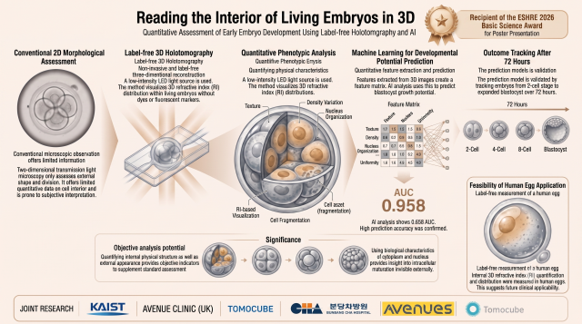
SEOUL, July 21 (AJP) - A South Korean research team has developed a way to look inside living eggs and embryos in three dimensions without damaging them, a technique fertility specialists say could sharpen how doctors choose which embryos to implant during in vitro fertilization.
The Korea Advanced Institute of Science and Technology (KAIST) said Tuesday that the work won the Basic Science Award for Poster Presentation at the European Society of Human Reproduction and Embryology (ESHRE) 2026 annual meeting, held in London from July 5 to 8. The prize goes each year to the single strongest basic-science poster at the world's largest reproductive medicine conference, which drew more than 10,000 participants. Judges shortlisted five finalists from the submitted abstracts, then decided the winner after live poster presentations and questioning.
The winning research came from a team led by Park Yong-keun, a physics professor at KAIST, with postdoctoral researcher Lee Chung-ha presenting the findings in London. The team paired time-lapse imaging, which follows living cells continuously without harming them, with a three-dimensional scanning method to predict early on whether an egg or embryo is likely to develop.
The success of IVF often comes down to picking the right embryo. Doctors now rely mainly on two-dimensional microscope images, weighing shape, size and how the cells divide to judge which embryos have the best chance of implanting. Because those embryos may later be transferred into a patient, doctors cannot use dyes or fluorescent markers that could alter or damage them, which leaves embryologists dependent on visual judgment calls that are hard to standardize.
Park's team turned to a technique called holotomography, which measures how light bends as it passes through a cell to build a three-dimensional map of its inner structure, all without staining or otherwise disturbing the living tissue. The method also captures the refractive index, a reading of how densely packed and what kind of material sits inside a cell, letting researchers spot fine internal differences that ordinary imaging misses.
Testing the approach on mouse embryos, the team found that embryos less likely to reach the blastocyst stage, the point at which a fertilized egg has divided enough to prepare for implantation in the uterus, tended to show a more uneven spread of refractive index inside their cytoplasm. Tracing that unevenness in mouse eggs, the researchers linked the brightest, highest-refractive areas to lipid droplets, the small fat-storing structures scattered through the cytoplasm.
The team then tried the same imaging and analysis on human eggs, not to predict how they would develop but to confirm the method could work at all beyond a mouse model. It did. The researchers captured three-dimensional refractive index maps of human eggs and put numbers to the same textural patterns, some of which again traced back to lipid droplets. But not every feature matched across species. Mouse and human eggs differ in size and internal makeup, so similar-looking optical signatures may not carry the same biological meaning. The researchers called the human trial a feasibility check rather than a proven predictive tool, and cautioned that markers developed in mice should not be applied directly to human eggs.
The project brought together KAIST's physics department, the fertility center at CHA Bundang Women's Medical Center under professor Kim Ji-hyang, the London fertility clinic Avenues and Tomocube, a holotomography equipment maker Park co-founded. Findings from the mouse embryo and mouse egg studies have been posted as preprints and are under peer review at international journals.
"This award matters because it recognizes the academic value of a new approach that lets us analyze living eggs and embryos in three dimensions, quantitatively, without damaging them," Park said. "We are now validating the method using human cells, and we will keep developing it into an objective, accurate evaluation technology that can help raise the success rate of infertility treatment."
(Reference Information)
Journal/Source: Preprint
Title: Label-free 3D subcellular phenotyping of mouse embryos by holotomography enables early prediction of blastocyst formation
Link/DOI: https://doi.org/10.1101/2024.05.07.592317
Journal/Source: Preprint
Title: Holotomography reveals biophysical remodeling of mouse oocytes during post-ovulatory aging
Link/DOI: https://doi.org/10.64898/2026.06.18.733271

Copyright ⓒ Aju Press All rights reserved.